
-
-
Koszyk jest pustyRealizuj rezerwacjęSuma Netto 0
- Produkt miesiąca
Cena po zalogowaniu
Do końca promocji pozostało
Cena po zalogowaniu
Do końca promocji pozostało
- Promowane produkty
Cena po zalogowaniu
Cena po zalogowaniu
Cena po zalogowaniu
Cena po zalogowaniu
Cena po zalogowaniu
Cena po zalogowaniu
Cena po zalogowaniu
Cena po zalogowaniu
- Nowości
Cena po zalogowaniu
Cena po zalogowaniu
Cena po zalogowaniu
Cena po zalogowaniu
Cena po zalogowaniu
Cena po zalogowaniu
Cena po zalogowaniu
Cena po zalogowaniu
Cena po zalogowaniu
Cena po zalogowaniu
- MEGAVET
Katalog Firm Weterynaryjnych
Od 2011 roku tworzymy największy Katalog Firm Weterynaryjnych w którym znajdują się najlepsze produkty i usługi współpracujących z nami producentów, dystrybutorów oraz usługodawców branży weterynaryjnej. W myśl zasady All-in-One oddajemy w Państwa ręce platformę weterynaryjną megavet.eu, której celem jest przedstawienie możliwie najszerszego portfolio do wyposażenia placówki weterynaryjnej "od A do Z".
Szukasz sprzętu weterynaryjnego?
Bez względu na to czy prowadzisz gabinet, lecznicę, przychodnię, klinikę czy szpital weterynaryjny platforma megavet.eu jest po to aby Ci pomóc zaoszczędzić Twój czas i pieniądze. Na naszej stronie, umożliwiamy zadawanie pytań zarówno o konkretne produkty weterynaryjne, jak również zapytania ogólne np. "Otwieram nową placówkę weterynaryjną. Proszę o wysłanie oferty na całe wyposażenie" - Zapytania ofertowe. Zapraszamy do składnia zapytań - to nic nie kosztuje, a wzbogaci Państwa o cenną wiedzę na temat poszukiwanych produktów bądź usług.
Reprezentujesz dostawcę, prodcenta lub usługodawcę?
Chcesz wypromować swoją firmę oraz jej portfolio? Chcesz otrzymywać bezpośrednie zapytania o swoje produkty lub usługi? Nie czekaj i skontaktuj się z nami - Kontakt. Omówimy najlepsze rozwiązanie dla Twojej firmy.
-
Warunkowy dostęp do
portalu megavet.eu
Dostęp do zawartości portalu megavet.eu jest możliwy wyłącznie dla osób wykonujących zawód medyczny lub prowadzących obrót wyrobami medycznymi.
Czy jesteś osobą zawodowo związaną
z branżą medyczną lub weterynaryjną?





![Analizator hematologiczny ProCyte Dx [IEX]](/images/megavet/3000-4000/Analizator-hematologiczny-ProCyte-D-_%5B3696%5D_260.jpg)
![URIT 11G wysokiej jakości paski do analizy moczu [MEO]](/images/megavet/19000-20000/ANALIZATOR-BIOCHEMICZNY-EMP-168-VET-MEO_%5B19777%5D_260.jpg)
![Analizator biochemiczny Catalyst One [IEX]](/images/megavet/3000-4000/Analizator-biochemiczny-Catalyst-One_%5B3695%5D_260.jpg)
![Cardell Insight ciśnieniomierz dla zwierząt [GWV]](/images/megavet/0-1000/Cardell-Insight-cisnieniomierz-dla-zwierzat_%5B972%5D_260.jpg)
![USG VINNO G65 VET [NRM]](/images/megavet/18000-19000/USG-VINNO-8-VET-NRM_%5B18346%5D_260.jpg)
![Sprzęt do zabiegów fizykalnych Viofor JPS [ALD]](/images/megavet/19000-20000/ACESO-80A-Automatyczny-analizator-immunochemiczny-ALD_%5B19204%5D_260.jpg)
![Analizator hematologiczny ProCyte One [IEX]](/images/megavet/18000-19000/Analizator-hematologiczny-ProCyte-D-IE-_%5B18245%5D_260.jpg)




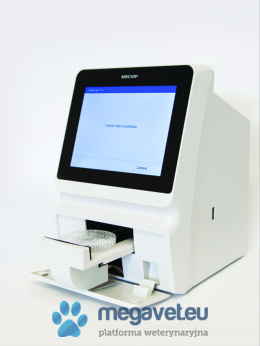
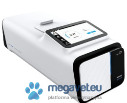
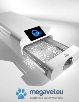
![ANALIZATOR DO HEMATOLOGII URIT 3000 PLUS [MEO]](/images/megavet/0-1000/ANALIZATOR-DO-HEMATOLOGII-URIT-3000-PLUS-MEO_%5B461%5D_260.jpg)
![Wideoendoskop weterynaryjny KARL STORZ 10,4 mm x 180 cm, kanał rob. 2,8 mm [MEM]](/images/megavet/10000-11000/Wideoendoskop-weterynaryjny-STORZ-10-4-mm-180-cm-kanal-rob-2-8-mm-MEM_%5B10817%5D_260.jpg)


